Akoya QPTIFF Slides in the Browser
Open the slides your Akoya PhenoCycler-Fusion or Vectra saves, as they are. Nothing to convert, nothing to upload. A large slide starts showing right away, because the viewer reads only the part you look at.
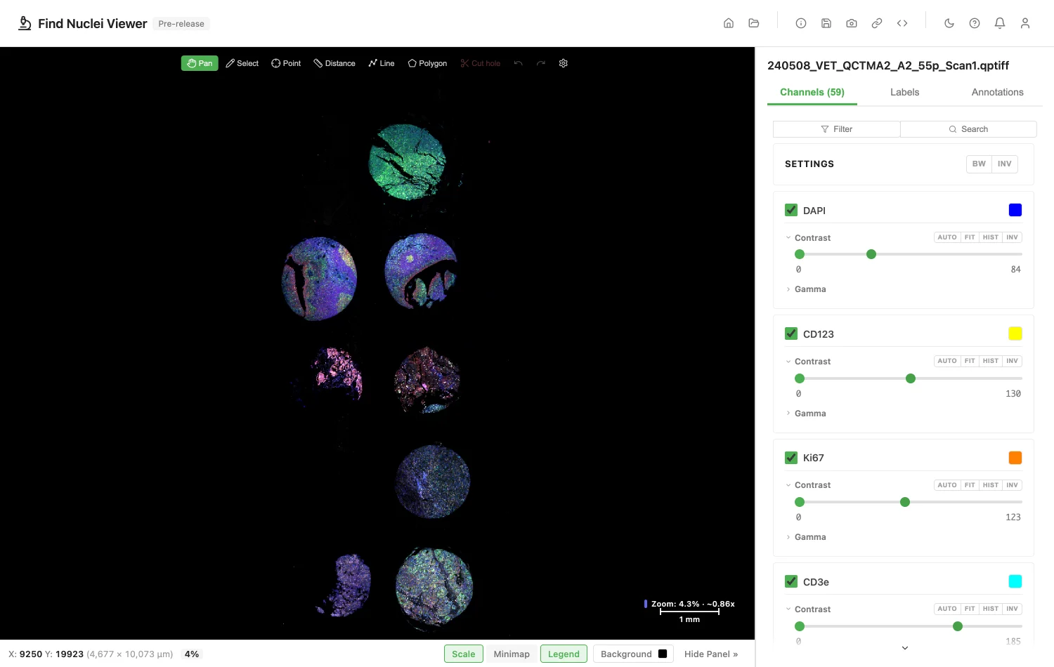
Multiplex fluorescence opens with every channel, named and coloured as in the scan.
How It Opens
- One file on your disk, in any browser. Drop it on the viewer, or click Open Image and Choose File. See Local Files.
- A folder of slides, in Chrome and Edge. One slide opens, several show a list. See Local Files.
- An address:
https://,s3://orgs://. Paste it under Open Image. See Remote URLs. - Private data, with a private link or a token. See Private Images and Slides.
What You Will See
- The slide. Zoom from the whole slide down to single cells.
- Brightfield. H&E and other stains open in colour.
- Multiplex fluorescence. Each channel opens with its marker name and colour from the scan, such as DAPI, CD3e or Pan-Cytokeratin. A scan of 59 markers opens with all 59, the first 10 turned on. Turn channels on and off, and set each one's contrast.
- A channel with no colour. Shown in white, with a note.
- Pixel size and magnification. Both come from the scan, so measurements read in µm and the zoom bar shows the objective. To set your own, see Pixel Size and Magnification.
The thumbnail, overview and label pictures in the file are not shown.
inForm Component Exports
inForm saves the unmixed result as a .tif file, one channel per component, such as Hematoxylin and Eosin. Drop it on the viewer like a slide.
It opens dark on white, so it reads like the stained slide it came from.
If a Slide Does Not Open
The viewer tells you why.
| What the file has | What the viewer says |
|---|---|
| Another compression | This QPTIFF uses a compression the viewer cannot read yet. |
| Another pixel type | This QPTIFF has a pixel type the viewer cannot read yet. |
| Pages of different pixel types | The pages of this QPTIFF do not share one pixel type. The viewer cannot read it yet. |
| Several planes per channel | This QPTIFF holds more than one plane per channel. The viewer reads one plane per channel for now. |
| A resolution missing a channel | A resolution level of this QPTIFF lacks a channel. The viewer cannot read it yet. |
| Not a QPTIFF | This file is not an Akoya QPTIFF. |
| Damaged or cut short | This QPTIFF looks damaged or incomplete. |
Annotations
Import GeoJSON or OME-XML, and export both, from the Annotations panel. Send a link that opens the slide with its annotations. Exchanging Annotations shows how. To draw, measure and label shapes on the slide, see Annotations.
Public Slides to Try
Zenodo record 15825670 holds a 59-marker PhenoCycler-Fusion scan of a tissue microarray, CC BY 4.0. Download the zip, unpack it, and drop the .qptiff on the viewer.
For Your IT Team
To open a slide by address, the server must answer byte-range requests and allow them through CORS. For a bucket, allow GET and the Range header in its CORS rules. What the Server Needs has the details.
When a server cannot send parts of a file, the viewer says so. Download the slide and open it from your computer.
Addresses that start with s3:// and gs:// work too.
Image credits
The screenshot uses 240508_VET_QCTMA2_A2_55p_Scan1.qptiff from Zenodo record 15825670, by Bryce Fukuda, Jocelyn Wright, Kimberly Smythe and Cecilia Yeung, raw data for the publication "Graphical and Interactive Spatial Proteomics Image Analysis Workflow", CC BY 4.0. See Sample Data and Credits.
Privacy
The slide is read in your browser. The file name and address are never recorded. See the privacy policy.
Product names are trademarks of their owners. Find Nuclei is not affiliated with them.